Showing posts with label bacteria. Show all posts

Friday, June 29, 2018

I've got the power: How the potential bioterrorism agent Francisella tularensis manipulates host cells


In an age of advanced weaponry and warfare, the risk of bioterrorism is increasingly acute. One potential bioterrorism agent is the bacterium Francisella tularensis, which is responsible for the similarly named disease tularemia. F. tularensis is classified as a category A potential bioterrorism agent, the same classification as anthrax and the plague. A low number of bacteria are capable of causing disease, which can be fatal in up to 60% of cases if untreated. Outside the potential threat for bioterrorism, F. tularensis infection also happens naturally. While cases of tularemia in the United States have largely declined since the 1950s, this is not the case throughout the world, with multiple outbreaks occurring in Europe in the last 10 years.
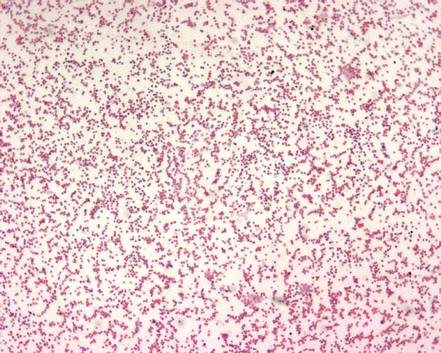
F. tularemia.
CDC's Public Health Image Library.
Image # 1903; photo credit: Larry Stauffer, 
Oregon State Public Health Laboratory.
Disease management and bacterial elimination can be difficult because F. tularensis can survive in over 100 species of mammals, birds, cold-blooded animals, and arthropods, including rabbits, mice, rats, squirrels, cats, dogs, horses, pigs, and sheep. To further complicate matters, transmission of F. tularensis can occur in several ways, including the consumption of contaminated water or food; contact with urine, excrement, or blood from infected animals; bites from blood-sucking arthropods like ticks, flies, and mosquitoes; and inhalation of aerosolized bacteria. The symptoms of tularemia depend on the route of transmission and can include a skin ulcer at the site of bacterial entry; swollen glands; sore throat; and high fever. F. tularensis is naturally resistant to many antibiotics because it is an intracellular bacterium that spends most of its life hiding inside a host cell; an antibiotic must first get into the host cell before it can have any effect on the pathogen. Aminoglycosides, tetracyclines, and fluoroquinolones have been shown to be effective, but 5-15% of infections relapse following treatment, and the side effects from these antibiotics can be unmanageable, limiting their use.

Due to the low infectious dose, high mortality rate, ease of transmission, and difficulty in treatment, natural F. tularensis infection is a serious threat to public health, and a weaponized version of the bacteria could be catastrophic. To counteract these risks, researchers have been studying how F. tularensis causes infection to identify ways to inhibit or kill the bacteria. We know that once in the human body, F. tularensis is taken up by phagocytic cells, such as macrophages. The job of these phagocytic cells is to engulf the bacterium into a compartment called a phagosome for degradation. Typically, this is how the immune system would capture and kill a pathogen. However, in the case of F. tularensis, the bacterium escapes from the phagosome through a process that is not well understood to begin replicating in the cytosol of the host cell. A recent study shed a little light on this process and found that F. tularensis is manipulating the host macrophage in a unique way.
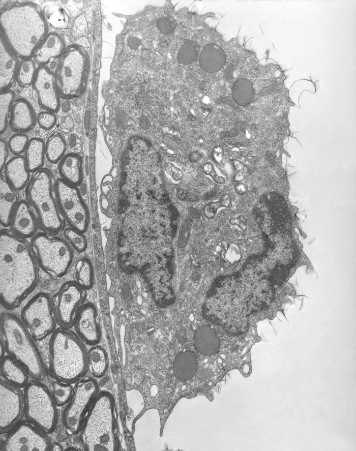
Macrophage (right) containing rickettsial microbes.
CDC's Public Health Image Library.
Image # 8731; photo credit: CDC, Dr. Ed Ewing.
Dr. Forrest Jessop and colleagues at the National Institute of Allergy and Infectious Disease found that F. tularensis alters the function of the mitochondria in the macrophage. The mitochondria are essential cellular organelles that are responsible for providing “power” to the cell, much like a battery provides power to a flashlight. When F. tularensis first enters the macrophage, it improves the function of the mitochondria, which keeps the macrophage alive and prevents an inflammatory response from the immune system. A few hours later, the bacterium reverses these effects and decreases mitochondrial function, decreasing the macrophage's power supply and leading to rapid bacterial replication and oncosis, a type of cell death that involves the swelling of the cell. This facilitates the pathogen’s ability to get out of the cell after replicating and move on to a new host cell.

The researchers were able to take this new-found knowledge of the bacteria’s effect on mitochondria a step further and test a therapeutic treatment in culture. They found that by treating F. tularensis-infected macrophages with drugs that protect typical mitochondrial function, they were able to reduce macrophage cell death and decrease levels of bacterial replication. It remains to be seen if this type of intervention will work in an animal system, but this is a promising step in the right direction towards increasing the number of treatments available for these infections. Since the environmental reservoir for F. tularensis is so vast, increased awareness of the risks of disease and research focus are important to stem the outbreaks and prevent future bioterrorism threats.

Thursday, May 31, 2018

Broadly neutralizing antibodies take down bacteria, viruses, and yeast

Antibodies are an important player in the body's immune system. The job of an antibody is to recognize a very specific feature of a foreign protein, known as an epitope. Without high levels of epitope specificity, antibodies can often begin to bind to and attack self-proteins, leading to dangerous autoimmune reactions. By producing highly specific antibodies, the body can avoid these autoimmune disasters. However, this high level of specificity also means that antibodies typically only recognize one specific species or even sub-species of foreign invaders. Interestingly, a research group recently identified antibodies from healthy individuals that could recognize multiple subgroups of Klebsiella pneumoniae, as well as various other bacteria and even some yeasts and viruses. These “universal antibodies” have sparked a great deal of interest as a potential treatment option for patients suffering from infections.

K. pneumoniae is a species of bacteria that is found frequently in the environment and in people; it is estimated that over 1/3 of the world’s population is colonized by K. pneumoniae. In individuals with a healthy immune system, the level of bacteria is controlled, and illness does not occur. However, in immunocompromised and already-ill people, the bacteria can cause severe infections. Klebsiella are the third leading cause of hospital-acquired infections in the United States, with alarmingly high mortality rates: K. pneumoniae pneumonia can cause mortality in up to 50% of patients, and bloodstream infections can cause mortality in 20-30%. Equally concerning is the rising level of antibiotic resistance found in K. pneumoniae. This makes them increasingly difficult to treat.


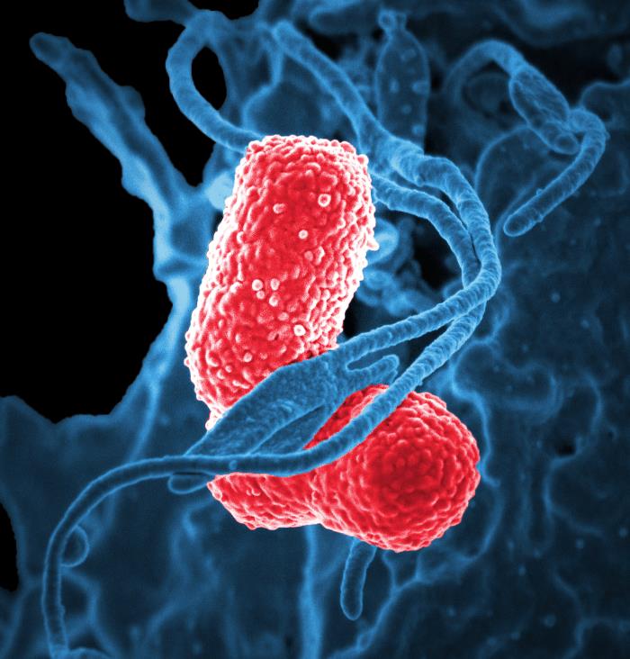
K. pneumoniae (red).
CDC's Public Health Image Library.
Image # 18170; photo credit: NIAID.
The outer membrane of K. pneumoniae, and other bacteria, is covered in lipopolysaccharide (LPS) molecules. Because this molecule is exposed to the external environment, LPS serves as a good target for the immune system to produce antibodies against. LPS is made up of repeating sugar residues; in the case of the K. pneumoniae subgroups of interest in this research, that sugar is mannose connected by 1-2 or 1-3 linakages. Researchers identified a number of antibodies from the blood of healthy individuals that were highly efficient at neutralizing K. pneumoniae bacteria by binding to these mannose residues. Because mannose is a common surface sugar molecule and the mannose molecule arrangements used by K. pneumoniae are also used by other microbes, these antibodies had a very broad specificity. They bound not only K. pneumoniae, but also other intestinal microbes, HIV virions, and the yeast Saccharomyces cerevisiae

These antibodies open the possibility for their use as therapeutics. Giving pre-made antibodies to patients has already been established as an effective strategy to treat or prevent a number of infectious diseases, such as rabies, diptheria, tetanus, hepatitis B, and botulism. In the case of these infections, antibodies that are highly specific to the pathogen of interest have been made and used. However, the identification of these broadly neutralizing antibodies opens the door for a new opportunity. Giving a patient "universal" antibodies could help fight a variety of infections without even necessarily identifying the causative agent, which can be difficult and time-consuming in the face of a life-threatening infection. While it is a long process from antibody identification to the approved use of an antibody as a therapy in patients, this discovery provides researchers direction for the path ahead. Therapeutic advances that use alternative strategies to inhibit and kill pathogens are of the utmost importance in the current age of antibiotic resistance. Antibodies, instead of just antibiotics, that can fight disease will be one of the important tools in our arsenal against the ever-evolving microbes.

Monday, July 31, 2017

New diagnostic method may help fight antibiotic resistance

Coughing, sneezing, runny nose, wheezing; respiratory tract infections are extremely common, with the average American adult enduring two to four each year. Typically, the symptoms last for seven to ten days while you struggle through, and then you get better. If you go to a doctor with these symptoms, they will likely prescribe you a broad-range antibiotic. This may sound fine, but there is a big issue with that: not all respiratory tract infections are caused by a bacterial infection. Over-use of antibiotics has been leading to increased antibiotic resistance for decades. The only way to prevent the over-prescribing of antibiotics for these respiratory tract infections is to determine the cause of each infection. Unfortunately, current technologies for diagnosing these infections are not fast and specific enough to allow timely and proper diagnosis. New technology has emerged that may help with the diagnosis and cut down on the over-use of antibiotics.

When exposed to different pathogens, the body's immune system responds in different ways. A virus, for example, causes cells to react in a different way than a bacterium. The cells of the immune system that can be found in the blood can be profiled to understand what type of infection they are fighting. This has been done by studying the messenger RNAs (mRNAs), also known as transcripts, found within monocytes, a specific subset of immune cells. mRNAs lead to the proteins being made by a cell and often play key roles in regulating the activation of pathways involved in an immune response. A recent study identified ten different mRNAs that could be used to determine if the body was responding to a bacterial or a viral respiratory tract infection. A more recent study further validated these ten mRNAs by confirming their use in 94 hospitalized adults with respiratory tract infections and identified even more mRNAs that could differentiate a bacterial infection from a viral infection. This allows for more appropriate use of antibiotics in these patients and avoids the potential over-use of antibiotics that threatens their effectiveness.

As more and more antibiotic-resistant infections emerge, it is becoming more important than ever to safeguard our potent antibiotics by only using them when necessary. When antibiotics are used, they kill off whatever bacteria are susceptible to their effects, leaving behind only those that are resistant. This helps select for antibiotic-resistant bacteria within a population. The over-use of antibiotics has sped this natural selection process, allowing for the rapid development of resistance even to the newest antibiotics. Using technologies such as this transcript profiling of immune cells will help slow the selection process by ensuring antibiotics are only introduced when they can be of help, allowing our antibiotics to maintain their usefulness longer before resistance develops.

The technology to profile transcripts of immune cells in the blood has the opportunity for application far beyond the identification of bacterial versus viral respiratory infection. Individual pathogens themselves can produce unique immune profiles that could one day be categorized using similar methods. With the standard diagnostics of the past, it is nearly impossible to diagnose a bacterial or viral infection unless the bacteria can be cultured or the virus can be isolated. Knowing the unique transcript profile induced by a pathogen could someday allow for molecular diagnosis of a number of pathogens without the need to culture the bacteria or isolate the virus, allowing for an even more significant reduction in antibiotic over-use. This would also provide more rapid diagnosis; transcript analysis could be performed in a matter of hours, while the diagnostics of the past frequently require days. With emerging technologies, these goals become even more achievable every day, and we may soon see a time when antibiotics are only used for infections confirmed to be caused by a susceptible bacteria.

Thursday, June 29, 2017

More vaccination victories needed in the meningitis fight

Vaccinations have been proven time and time again to prevent disease and improve health outcomes. All around the world, vaccines have been deployed to deal with illnesses as common as the flu and as deadly as Ebola. Meningitis is another disease for which vaccination has become a major priority. The “kissing disease,” at it is sometimes called, has made a number of appearances on college campuses across the United States. While incidence in the U.S. remains quite low, at 0.3-4 cases per 100,000 persons, incidence can be as high as 1 case per 100 persons in the “meningitis belt” of Africa, where epidemics occur with regularity.

Image result for neisseria meningitidis
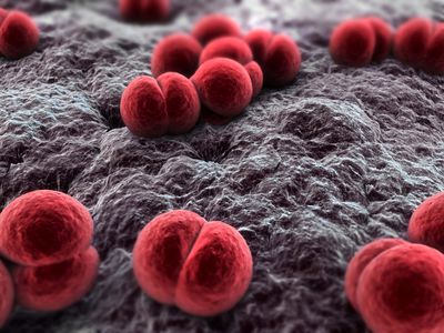
Neisseria meningitidis, the bacterium responsible for meningitis.
Image from Bioquell.com
Infection with the bacterium Neisseria meningitidis, the major cause of meningitis, often goes unnoticed. The bacteria take up residence within the nasal cavity, where they can stay without causing disease in a carrier individual. However, in approximately 1-5% of people exposed to the bacteria, invasive disease occurs, and the bacteria enter the bloodstream, leading to life-threatening disease.

Symptoms of meningitis typically begin almost immediately, just one day after infection, and include flu-like symptoms of fever, headache, and stiffness. Because the bacteria enter the bloodstream, any organ or tissue can become infected and impaired. Despite years of research, mortality rates continue to range from 10-15%, even in developed countries, with rates above 20% in the developing world. Even for those who survive the invasive disease stage, meningitis causes lasting impairments in 19% of patients, with neurological disabilities, seizures, hearing or visual loss, and cognitive impairment being classical manifestations. The rapidity of disease progression, along with the high mortality rate, make meningitis a prime disease target for vaccination.

The first vaccines against meningitis were developed in the 1970s. Unfortunately, these early vaccines lacked the ability to maintain long-lasting immunity against the bacteria. In the late 1990s, alterations were made in the vaccine components, allowing for the elicitation of an immunological memory response that would be effective to protect young children into their adult years and would even help reduce the rates of carriage of the bacteria in the nasal cavity. While this was great news for the prevention of meningitis, challenges still remained. The bacteria that cause disease can belong to any of 6 different serogroups, meaning that immunity to one serogroup will not necessarily provide protection from another. This requires differential targeting of all 6 serogroups to truly prevent disease.

Image result for meningitis vaccine
Image from the Meningitis Vaccine Project
Researchers have addressed this challenge by producing different vaccines for use in specific parts of the world where each serogroup is problematic. In the meningitis belt of Africa, for example, serogroup A has historically been the cause of epidemics. To wipe out these epidemics, a mass vaccination campaign was begun in 2010; the Meningitis Vaccine Project produced and provided vaccines against N. meningitidis serogroup A for over 217 million people in 17 different countries. Thanks to these vaccines, epidemics linked to the serogroup A bacteria have been eliminated.

Unfortunately, when one serogroup is removed, a niche opens up for another. Just last month, the CDC announced that a small epidemic in Liberia had been caused by the N. meningitidis serogroup C bacteria. Nigeria and Niger have also reported outbreaks of this serogroup. Luckily, in the case of Liberia, the country’s response time was extremely rapid. Thanks to the health system improvements made during the Ebola outbreak, Liberia now has a robust case detection and monitoring system. Other countries in the area, however, are not nearly as advanced and could suffer a severe epidemic if serogroup C moves in with force.


Great strides have been made in the fight against meningitis outbreaks. However, the complexity of the group of bacteria responsible for the disease leaves a number of challenges in place that must be overcome. The ideal solution would be the introduction of a vaccine that combined pieces from each bacteria serogroup to produce an immune response in patients that would protect from all six serogroups at the same time. While some quadrivalent vaccines already exist, which provide protection against four of the six serogroups, these vaccines have only been recommended for use in the U.S. for adolescents entering college. Protection from this vaccine only lasts 2-5 years in adults, making it less than ideal for deployment in rural areas where boosting is not a viable option, such as Africa. Advances in vaccine technology may help improve the longevity of protection, making multivalent vaccination a more robust solution to the meningitis problem. Until then, rapid case detection and monitoring capabilities, such as those displayed in Liberia, will be the key to keeping meningitis epidemics in check as they arise. Between vaccine and monitoring advances, meningitis epidemics may one day become a thing of the past. 

Sunday, May 21, 2017

The Stressed-out Microbiome

Stress. It's something we encounter every day. People teach workshops and write books on how to deal with it. We spend countless hours and dollars trying to avoid it. But it's still there. Stress is as much a constant in our world today as the air around us. We all know that stress can have negative impacts on mental well-being, but we are just beginning to realize the effects stress can have on other bodily systems, and the effect these systems can have on our ability to deal with stress. Recent research has shown that the microbiome can be significantly affected by and play a major role in our response to stress.

The microbiome is the group of microbes (bacteria, fungi, archaea, etc.) that colonize a particular area of the body. The gut microbiome has been a population of intense research for quite a few years now. With increasing technology, we've begun to learn more and more about this population. We know that at homeostasis, each person has a specific microbial population that makes up their gut microbiome. This helps keep us healthy, keeping our digestive system running smoothly and ensuring successful processing of the food we eat. But in times of disease or stress, the microbiome in the gut can change dramatically, causing symptoms from loss of appetite to ulcers.

How exactly does stress change what's living in your intestines? It's all because of the gut-brain communications. This is achieved through neural projection pathways, neuroendocrine signaling, and entero-endocrine signaling, among other pathways. Your body uses messaging through the neurons and small chemical messengers in the form of endocrine signals to communicate over long distances, such as from the brain to the gut. In return, the gut uses the same mechanisms to send feedback to the brain. The microbes in the gut, however, can also produce chemical messages that get sent to the brain. When your body experiences stress, different chemical signals are sent to the gut; this can, in turn, cause some microbes to die and allow other microbes to take their place. The change in the composition of microbes can then change the chemical signals sent back to the brain. In this way, there is an intimate connection between the signalling input from the brain and the feedback the brain receives. These signals are essential to support mood, higher cognitive function, and behavior.

Work in animal models has highlighted the influence of stress on the gut microbiome and the subsequent influence of the microbiome on behaviors. For example, when mice are raised in a germ-free environment (no microbiome), the mice show reduced anxiety-like behaviors as compared to mice that are not raised in a germ-free environment. It has also been shown that the microbiome of the mice has an influence on the development of the amygdala, a region of the brain that plays a vital role in controlling behavioral and physiological responses to stress stimuli. The germ-free mice showed increased amygdala volume, suggesting an increased ability to handle stress. Additional studies have found that early-life stress can change an animal's microbiome for the rest of its life and that microbiome transplantation from depressed rats to healthy rats can lead to anxiety-like behaviors in the healthy rats.

All the interest in the microbiome and stress has led to new insights for the treatment of stress-related behaviors through microbiome alteration. Allowing colonization with particular species of bacteria, for example Lactoacillus helveticus and Bifdobacterium longum in both rats and humans, has been shown to reduce psychological distress. Additionally, treatment with oligosaccharides, such as fructo-oligosaccharide or galacto-oligosaccharide in mice, can have anti-depressant effects by allowing different populations of microbes to thrive on these nutrient sources.

While the microbiome still holds many mysteries, we are beginning to understand just how important its functions are in our every day lives. So the next time you're feeling stressed out, you might want to check in with your microbes and see how they're feeling. They may play a critical role in getting you through this stress and on with your life without allowing long-term health complications, like depression, anxiety disorders, or inflammatory bowel disease, to develop. The best ways to keep a healthy microbiome are to eat a healthy diet, with emphasis on fibers that support the development of beneficial bacteria, take probiotics, stay physically active, and avoid antibiotics whenever possible. Or, simply change your lifestyle to reduce your stress level. However it is achieved, a healthy microbiome can be a great advantage in the years ahead.

Wednesday, January 25, 2017

More diarrheal disease plagues The Oregon Trail

"James has died of dysentery." Another common diarrheal disease you likely remember from The Oregon Trail, dysentery is caused by the consumption of contaminated water, like cholera and typhoid. There are actually two different forms of dysentery; one form is caused by Shigella bacteria (also known as shigellosis), and the other is caused by Entamoeba histolytica, an amoebic parasite. E. histolytica causes milder disease, but is more difficult to treat than that caused by the Shigella bacteria. No matter which form of dysentery you have, the symptoms can include anything from mild diarrhea to severe, bloody diarrhea with a fever, cramps, vomiting, and even pain and complications outside the intestines.

Dysentery has been known as a disease for quite some time, with case reports dating back to the 1200s. In light of the recent presidential inauguration in the United States, it is interesting to note that one of our own U.S. presidents suffered from dysentery on the very day of his inauguration. In the weeks leading up to James Buchanan's inauguration in 1857, he came down with dysentery. He spent the two weeks before March 4, inauguration day, in seclusion in an attempt to be well enough for the ceremony. He even turned down a dinner invitation from the famed Jefferson Davis in a letter in which Buchanan said he was "now living with great caution." Buchanan was well enough by March 4 to be successfully sworn in without incident.

While dysentery is considered a fairly rare disease in modern times, it is still estimated that there are 170 million cases each year, along with 14,000 deaths. A recent increase of cases in Zimbabwe has caught much public attention. Corruption within the country has led to a suspected misuse of funds within the government. The Movement for Democratic Change party (MDC), which currently holds the presidency and other governmental positions, has been accused of using the country's money to buy cars and fund their 2018 political campaign instead of building infrastructure and dealing with water sanitation issues. Because proper water sanitation is essential for preventing a disease like dysentery, critics of the government believe the members of the MDC are responsible for the current cases of both typhoid and dysentery that have been spreading through Zimbabwe.

Although dysentery has existed for hundreds of years, our approach to treatment now is not very different from times gone by. Typically, dysentery clears by itself, so the only remedy is to provide a patient with fluids to prevent dehydration. While antibiotics can be prescribed for dysentery caused by Shigella, instances of antibiotic resistance in Shigella have been on the rise since the 1940s, making many classes of drugs ineffective. Recent work has found that probiotics may play a role in helping to reduce disease duration and intensity in Shigella dysentery, but further work will be required before probiotics can be used for patient treatment.. Treatment of amoebic dysentery, on the other hand, is still almost purely reliant on simply relieving the symptoms.

As has been highlighted in the cases of cholera, typhoid, and dysentery, sanitary drinking water is the key for the prevention of disease. While many countries are focusing efforts on improving water sanitation, it is a long road to improve an entire country's infrastructure enough to provide all residents with clean water. But support for these initiatives continues to pour in from other countries and private foundations alike, keeping alive the hope for a future with clean drinking water for all.

Wednesday, November 30, 2016

Further down the trail....Mary has died of typhoid

"Mary has died of typhoid." Yet another disease you may remember encountering on The Oregon Trail in childhood, typhoid fever, or simply typhoid, is also still a major public health concern today. Affecting an estimated 20.6 million people and causing 223,000 deaths a year, typhoid is caused by the bacterium Salmonella enterica serovar Typhi (S. Typhi). The bacteria is often spread through contaminated water or from person-to-person contact. In places where clean water and sanitation are standard, the disease has been essentially eliminated. But in developing countries, it remains a major threat.

The most recent outbreak of typhoid occurred just last week in the city of Masvingo, Zimbabwe. Areas where the disease is endemic and can lead to outbreaks include Southeast Asia, Africa, and South America.  Travel to and from these areas also allows for the transmission of the bacteria to industrialized nations and can cause local outbreaks. Although there are currently two approved vaccines against S. Typhi, both have drawbacks that prevent them from being used en masse. The protective efficacy of the vaccines is sub-optimal and ranges from 40%-70%, largely dependent upon age and location. Additionally, the protection from the vaccines is short-lived, averaging 2-3 years for one vaccine and 5-7 years for the other. A further complication is that both vaccines need to be stored with refrigeration until they are used. Transport and storage under these conditions are major difficulties in countries where electricity is a supreme luxury.

Without a reliable vaccine, the major way to fight the disease is through the use of antibiotics. Chloramphenicol, ampicillin, and trimethoprim-sulfamethoxazole, very common antibiotics, have historically been used to stop the infection, and this strategy was highly successful for many years. However, in recent years, a new threat has emerged to thwart these efforts. In the 1970s, cases of S. Typhi that were resistant to these antibiotics began to emerge. We now face the threat of multi-drug resistant S. Typhi, making disease treatment much more difficult.

In light of the rising drug resistance, many have begun to see wide-spread vaccination as the best strategy to fight typhoid. Others argue that improving water sanitation will have the greatest effect on decreasing disease prevalence. One thing is certain: either strategy will require a large investment of funds to be achieved.

The Bill & Melinda Gates Foundation is leading the way in the funding arena and has identified the elimination of typhoid as a public health problem by 2035 as a goal. The Gates Foundation recently gave over half a million dollars to Yale University to explore and determine the cost-effectiveness of typhoid vaccination strategies. Additionally, The Gates Foundation gave a $36.9 million grant earlier this month to a collaboration between the Maryland School of Medicine Center for Vaccine Development, the Oxford Vaccine Group, and PATH, a non-profit public health organization, to accelerate the development of a new vaccine to be used in young children. The goal is to develop a vaccine with more long-lasting protection than the two currently available. The Gates Foundation is also providing funds to increase surveillance for typhoid in South Asia and Africa.

While the Gates Foundation is leading the way to fund the vaccination strategy, other groups are focusing on improving water sanitation. The United States Agency for International Development (USAID) features water and sanitation as one of its main avenues of focus, with sponsored projects underway in large portions of Africa and Southeast Asia. The World Bank has also been sponsoring projects to improve water quality and health throughout the world. Additionally, UNICEF has been a major player in the increased access to clean water that has over the past decade. Many other smaller non-profits have also played a role in this endeavor.

As both vaccine and water and sanitation improvements occur, our ability to battle this age-old pathogen will increase. Using both strategies simultaneously allows for the most rapid and sustainable progress toward S. Typhi elimination. With the continued investment of groups like the Gates Foundation and USAID, the goal of typhoid elimination as a public health problem by 2035 might just be achievable.

Sunday, October 30, 2016

Cholera: Not just a problem on The Oregon Trail

"Sally has died of cholera." This was a common problem in the game The Oregon Trail that many people remember from childhood. Tragically, cholera is still a major public health problem in countries around the world today. Cholera is caused by the bacterium Vibrio cholerae, which is shed in fecal matter from an infected individual and is often transmitted via contamination of water sources. In countries with poor sanitation and a lack of clean water, this can lead to significant and deadly outbreaks.

Fast-forward from the days of the Oregon Trail to present-day Haiti. The country has suffered multiple tragedies in recent years. In 2010, they were rocked with a devastating earthquake; now, in 2016, they suffered the wrath of Hurricane Matthew. In light of these recent disasters, there have been humanitarian efforts from the United Nations and other relief organizations. Unfortunately, in the wake of the U.N.'s help after the 2010 earthquake, Haiti experienced its first cholera outbreak.

Before 2010, the small nation of Haiti had not been exposed to cholera. The citizens had not had the disease, and no immunity to the pathogen existed there. The first case of cholera was reported in mid-October 2010 in the region of the country along the Meille River. The disease quickly became rampant, with a hospital 60 miles away from the first case reporting new cases every 3.5 minutes within just 2 days. Since then, the outbreak has affected nearly 800,000 people and caused more than 9,000 deaths according to the official numbers; however, many experts fear the true impact has been far greater due to poor case reporting.

Flooding and destruction from Hurricane Matthew on October 4, 2016, have done nothing to help the situation. As many Haitians have lost their homes and their sources of fresh water, cholera has been on the rise again. Flooding has led to increases in contaminated waterways, leaving much of the water unsafe for consumption. Within four days of the storm passing through, officials were reporting 62 cases and 13 deaths from cholera.

The issue of how cholera came to be endemic in Haiti has been a topic of heated debate in the past few years. Many have blamed the U.N.'s Nepalese peacekeeping troops for bringing the bacteria with them into the country following the 2010 earthquake. The U.N. has denied any potential responsibility for the outbreak for years, even in the face of lawsuits from families of those who had died. Others hypothesized that increases in the temperature and salinity of the rivers throughout Haiti had allowed bacteria that may have been living in a dormant state in coastal waters to populate these rivers after the earthquake.

Scientific evidence, however, has been on the side of those who blame the U.N.'s peacekeeping troops for the introduction of the disease. Genetic analyses by whole genome sequencing of the bacteria found in Haiti in 2010 showed that this strain was highly similar to the strain found in Nepal in 2010. Additional studies using multiple-locus variable number tandem repeat analysis (aka DNA fingerprinting), a technique that looks at the number of times a DNA sequence is repeated at specific loci in the genome, also suggested a match between the Nepalese and Haitian strains.

On August 18, 2016, after 6 years of denial, the U.N. finally acknowledged that they did play a role in the initial outbreak. Farhan Haq, the deputy spokesman for the U.N. secretary general, said "over the past year, the U.N. has become convinced that it needs to do much more regarding its own involvement in the initial outbreak and the suffering of those affected by cholera...[a] new response will be presented publicly within the next two months."

The latest chapter in this story comes with the announcement on October 24 that the U.N. is working on a plan to spend about $400 million on cholera in Haiti. Roughly $200 million will be spent on cholera elimination efforts, while the other $200 million will be given directly to families or communities affected by the disease. The full details of the plan are expected to be solidified in the coming weeks. However, questions remain over how the money for this plan will materialize. U.N. member states have already expressed discomfort with paying money to directly compensate victims, as this is not within the purview of the normal development work the U.N. is chartered to perform.

While the final details of the U.N.'s action plan remain to be worked out, it looks like Haiti will be receiving some much-needed support to aid in their cholera elimination efforts in the near future. As the country rebuilds after Hurricane Matthew, water sanitation will be a major focus for the nation. With financial support and help from the U.N., experts are hopeful that cholera can be eliminated fairly quickly from Haiti. That would serve as a major beacon of hope for a nation that has borne the brunt of tragedies for too long.

Tuesday, August 16, 2016

Amanda Elmore and Team USA's victory in the Olympic W8+ is cause for excitement, but the health risks they faced to achieve this feat are not

Rio de Janeiro. Already well known for its vibrant culture and nightlife, this Brazilian city has also become known for sports this month as they play host to the Games of the 31st Olympiad. Many brand new athletic facilities were created specifically for these games, and Brazil poured a projected $18 billion or more into bringing these games to life. Even more funding was supplied by sponsors, like Coca Cola. Unfortunately, not all sports venues could be made ideal.

Guanabara Bay and Copacabana Beach off the coast of Rio are the sites of five aquatic events in this year's games: sailing, rowing, canoe sprinting, the triathlon, and marathon swimming. In addition to hosting these great sporting events, these waterways also play host to many unwelcome guests: multi-drug resistant bacteria and viruses of many varieties.

The Brazilian government has been aware for years that raw sewage has rushed into their waterways from the cities. In their bid to bring the Olympics to Rio, Brazil pledged to put forth $4 billion to deal with their water contamination issues. Unfortunately, due to a "budget crisis" they were only able to invest $170 million before the Games began. The results of this lack of funding may end up having devastating effects on the health of athletes at these games.

A study published by Renata Cristina Picao's group in Brazil in 2015 looked specifically at the bacterial populations of the water from the beaches surrounding Rio de Janeiro. They studied a total of 18 water samples from different regions along the coastline. Of these isolates, only one had bacteria with susceptibility to imipenem, a common drug used to treat bacterial infections in this area. Resistance rates to other popular drugs were also alarmingly high, with 77.8% of the isolates showing bacteria with resistance to cefotaxime, 50% showing resistance to cefepime, 27.8% showing resistance to gentamicin and amikacin, and 5.6% showing resistance to ciprofloxacin. With drug resistance running rampant in the bacteria that call this water home, being on or, even worse, in this water may pose a significant health threat to athletes.

Possibly an even larger threat than the bacteria in these waters are the viruses that can also be found. Hepatitis A virus can be found in human waste, and experts speculate that ~60% of Brazilian adults are exposed to the virus. In waters that contain large amounts of human waste, like the ones the athletes will be exposed to, the risk of infection is significant. The CDC recommends that all travelers to Brazil, not just those who will be exposed to the water, receive the Hepatitis A vaccine. With appropriate use of the vaccine, an outbreak of Hepatitis A can likely be prevented, though some have questioned whether or not the vaccine will protect against the local strains of the virus.

In addition to Hepatitis A virus, water tests have found alarmingly high levels of multiple types of adenovirus, which can cause severe gastrointestinal problems and do not have vaccines. Fernando Spilki, a Brazilian virologist, performed water testing for the Associated Press and found levels of adenovirus from 14 million to 1.7 billion virions per liter of water. To put this in perspective, California officials become concerned about their water quality if the level rises to just 1,000 virions per liter.

Many may wonder why, with the popularity of these beaches among Brazilians, there has not been a major viral outbreak or an outbreak of multi-drug resistant bacteria in the region already. The answer likely lies in the fact that these native-born and raised Brazilians have been exposed to these bacteria and viruses from a very young age, allowing their bodies to develop a successful immune response to the pathogens. However, the same immunity will not exist for the foreign athletes who will be exposed to these waters.

In the year leading up to these games, some athletic groups have already trained and raced on these waters. Many documented athletes experiencing illness. The World Junior Rowing Championships were held in Rio in 2015, and the U.S. team documented 13 rowers who suffered gastrointestinal illness following the event. The Australian sailing team has trained on the waters around Rio for the past several months, and they also have had athletes fall ill with gastrointestinal problems.

Though independent water testing has identified the water as potentially hazardous to health, the International Olympic Committee has maintained that the water is safe enough for the events to be held. The World Health Organization (WHO) has recognized that the water quality is less-than-ideal, and has issued several statements for travelers warning them of the potential for infection if exposure to contaminated water occurs. Additionally, the WHO has recognized that sites in the Guanabarra Bay, where sailing, rowing, and canoe sprinting take place, do not always meet the standards of safety, based on bacterial testing. As a precaution, they recommend that for all bodies of water "all athletes should cover cuts and grazes with waterproof plasters prior to exposure, try to avoid swallowing the water, wash/shower as soon as possible after exposure and, as far as possible, minimize their time in the water and avoid going in the water after heavy rainfall if possible." In events like the triathlon, where the swim portion is upwards of 20 minutes, and the marathon swim, which can take 2 hours or more to complete, minimizing time in the water is not always a viable option.

In light of the potential health risks, athletes and spectators alike will need to use increased caution regarding the Rio games. Monitoring for illness is going to be critical to prevent severe illnesses from developing. Many have focused on the potential threat of Zika virus at these games, but the threat of the multi-drug resistant bacteria and viruses in the waters should not be forgotten. All can cause significant problems and really ruin the Olympic experience. Perhaps next time the Olympic Committee will be more skeptical of selecting a location with such serious health concerns unless they are willing to chip in some funds to help the country address the situation. Such a gesture would not only have a positive impact on the athletes, but more importantly, it would have a sustained impact on the residents of the host country for years after the conclusion of the Games.


--On a side note, I'm so proud of my former teammate Amanda Elmore and the entire U.S. W8+ for dominating and winning gold at Rio!! Boiler Up!

Monday, July 18, 2016

The wonders of the biofilm world

What do you moving your arm and a biofilm of bacteria growing have in common? The answer is more than you might think. You moving your arm involves the propagation of an action potential through neurons that connect your brain with your limbs. This action potential is based on the rapid movement of ions into and out of cells, allowing each cell to pass a message on to the cell next to it through these ions. As the charged ions flux in and out of cells, the membrane potential (or chemical voltage) of the cells changes. It turns out that biofilm bacteria can use a similar system in order to communicate.

from Anatomy & Physiology by Phil Schatz
Action potentials have long been known as a rapid way to propagate signals over long distances. As ion channels open or close over the course of the action potential, the charged particles flow in and out of the cell in response to their concentration gradients. This is what allows the changes in membrane potential within the cells. But it has only recently been found that Eukaryotes are not the only organisms that can do this.

Enter Bacillus subtilis, a bacterium often used as a model organism for studying biofilms. A biofilm is a collection of bacteria that adhere to each other and, often, a surface. Biofilms are more resistant to antibiotic treatment than free-living bacteria, and can commonly be formed on medical devices, such as catheters. It has been known for many years that bacteria within a biofilm are able to communicate through a process known as quorum sensing, which involves the release of chemicals by the members of the biofilm to control the population density. Recently, a study found that in addition to quorum sensing, B. subtilis cells can communicate through the creation of and propagation of action potentials, similar to neuronal signaling.

It was observed that the entirety of the B. subtilis biofilm would undergo metabolic changes in response to glutamate and ammonium nutrient limitation affecting the cells in the center of the biofilm. In order for these widespread metabolic changes to occur, the cells in the interior of the biofilm must communicate with the cells of the periphery. It was found that an active propagation of a potassium ion signal through the use of potassium channels on the cells was responsible for this communication. As the potassium channels on the surface of the bacteria opened, potassium would rush into the cells from the surrounding environment, resulting in membrane depolarization. The membrane depolarization is linked to a decreased ability of the cells to take up glutamate and ammonium, allowing these nutrients to build up and replenish the supply to the interior cells.

Biofilms are notoriously difficult to treat when they form in patients. As many as 80% of chronic infections are caused by biofilm formation. Persistent staphylococcal infections are often caused by biofilms, as are Pseudomonas aeruginosa lung infections. It is typically the interior cells of the biofilm, which have a more dormant lifestyle, that are most responsible for antibiotic resistance. Learning more about how the biofilms communicate can facilitate improved treatment. If the action potential creation of these bacteria can be inhibited, the cells will be unable to communicate in times of nutrient depletion, leading the cell death at the interior of the biofilm. This could greatly improve the ability to treat these infections, leading to better outcomes for patients. Perhaps some day soon, we will have better tools for treatment to gain ground against these crafty biofilm bacteria.

Beyond the impact of these findings on patients, it is truly marvelous to see what these relatively simple organisms can accomplish. Bacterial cells are almost 1000 times simpler than mammalian cells when genome sizes are compared, yet they are capable of accomplishing signaling akin to the complexity of neuronal signaling. There seems to be no end to the surprises these timeless organisms have in store for us. Who knows what will come to light next.

Thursday, March 31, 2016

A new leader in minimalistic genomes is born

Thanks to the work of scientists at the J. Craig Venter Institute in San Diego, CA, a new minimalistic microbe has been brought into the world. Through genome engineering, they have created a synthetic bacterium called Syn 3.0 that requires only 531,000 bases in its genome to grow with a doubling time of 3 hours in the laboratory. The next smallest free-living organism, Mycoplasma genitalium, has a genome of 600,000 bases, but grows with a doubling time of about 2 weeks. For comparison, consider the more well-known bacterium, Escherichia coli, which has a genome of 4,639,221 bases and a replication time of about 30 minutes in the laboratory. But what makes this new organism, which is approximately one-ninth the size of E. coli’s genome, able to survive and grow so readily? This question is especially puzzling in light of the fact that approximately one-third of Syn 3.0's genome codes for genes of unknown function.

Ever since the invention of genome sequencing, scientists have been identifying genes of unknown function. Even in the most well-studied of organisms, like the mouse, almost 96% of the genome remains of unknown function. Many of these segments are considered important for higher organization of the genome, allowing tight regulation of expression of the genes that code for specific RNAs and proteins. Bacteria tend to have the most completely annotated genomes of the model organisms due to their simplicity. In E. coli, 66% of the genes are of known function, and as much as 76% of the genome can be assigned a function by biochemical analysis software. Since approximately 3 million of E. coli’s 4 million bases of genetic material have known functions, it is rather shocking to find another bacterium that contains so many segments with unknown functions. Since Syn 3.0 has the smallest genome the researchers at the Venter Institute could engineer that could successfully sustain life, this suggests that we still do not know the functions of many essential genes.

In order to identify the function of genes of unknown function, many approaches can and have traditionally been used by researchers. The oldest method is to use random mutagenesis. Through this technique, you are able to use chemical mutagens or electromagnetic radiation to induce changes in different bases throughout the genome. After mutagenesis, you can identify what processes the organism can no longer perform. Sequencing can allow you to identify where the mutations you introduced occurred, thus helping link those genes with a molecular process. If you are only interested in one molecular process, you can design a screen to specifically pick out mutants that are deficient in this process for analysis.

More recent advances in genetic engineering have allowed for more sophisticated analyses. You can now delete a specific gene of interest and observe the phenotype. Alternatively, you can tag a gene with a marker, so that the specific protein produced is linked to a fluorophore or tag. This allows you to identify where and when the protein is expressed. The advent of new genetic engineering technologies, which made production of Syn 3.0 possible, will also enable us to discover the functions of those unknown genes.

The invention of this novel minimalistic microbe shines new light on our true lack of understanding of genetic material in organisms. By improving our knowledge of this unique bacterium, we can hope to improve our understanding of our own genome, and that of our many pathogens. Discoveries made from Syn 3.0 may be the key to great steps forward in understanding the genetic basis of disease and finding cures for the future.

Sunday, May 10, 2015

A look at the epidemiology of carbapenem-resistant Enterobacteriaceae

This piece is adapted from a paper I wrote recently for an emerging infectious disease epidemiology class.


Carbapenem-resistant Enterobacteriaceae: An Emerging Threat
Introduction to Enterobacteriaceae
The bacterial family Enterobacteriaceae is a diverse family that contains many of the members of the typical gut microbiota, including the potentially pathogenic Klebsiella pneumoniae and Escherichia coli, among others (1). This family has a long history of causing infectious disease when these bacteria make their way to sites other than the gastrointestinal tract. The advent of antibiotics, however, put a sharp end to that history. Since the introduction of penicillin in 1945, the incidence of these infections has decreased drastically (2). Unfortunately, in recent years there has been an emergence of carbapenem-resistant Enterobacteriaceae that are now threatening to un-do the strides that have been made in elimination of these infections.

Enterobacteriaceae is a family of gram-negative, rod-shaped bacteria. They can cause a variety of disease manifestations, depending upon the body system infected. Symptoms can include, but are not limited to, fevers, pain and/or pus from wounds, and severe pneumonia (2). These infections are typically diagnosed through laboratory analysis to isolate the organism from blood, urine, or cerebrospinal fluid (2). Typical Enterobacteriaceae infections are easy to treat with a class of antibiotics known as the carbapenems. The carbapenems include drugs such as ertapenem, meropenem, and imipenem. They are bactericidal members of the β-lactam family (3), the same family as penicillin, and they have a long track record of successful clearance of infections.

Carbapenem resistance and the impact on disease
Carbapenem resistance among Enterobacteriaceae in the United States and the world was almost non-existent before 1992. In the time from 1986 to 1990, the National Nosocomial Infection Surveillance system reported that 2.3% of the Enterobacteriaceae samples tested were deemed “not susceptible” (4). It was known during that time that many of these bacteria possessed extended-spectrum β-lactamases that were capable of degrading many other antibiotics and giving them the distinction of “not susceptible,” but they were still susceptible to the carbapenems (5). Since then, carbapenem resistance has been on the rise. Using isolates submitted for the Meropenem Yearly Susceptibility Test Information Collection Program, the incidence of resistance by different bacteria to these carbapenems has been tracked. Among K. pneumoniae alone, the incidence of resistance to meropenem increased from 0% in 1999 to 5.6% in 2008. The trend for E. coli was slightly less worrisome, with the incidence of resistance to meropenem and imipenem rising to just 0.8% and 0.2% respectively (6). With the emergence of resistance to the carbapenems, there are highly limited therapeutic options remaining to fight these infections (1). Most carbapenem-resistant isolates show resistance to all standard antibiotics. The drug of choice for these infections has typically become colistin. Alarmingly, however, cases of K. pneumoniae that were resistant to both carbapenems and colisin were found in 2009 in three different medical centers in Detroit, MI (7). Other potential treatment options, such as polymyxin and tigecycline, remain experimental at this time (8).

The increase in drug resistance among these pathogens has led to an analogous increase in disease. These pathogens are typically acquired in the hospital setting, so this is where surveillance generally takes place. Risk factors for infection include advanced age, increased illness severity, increased length of hospital or intensive care unit stay, the use of catheters, ventilators, dialysis, surgery, and prior exposure to β-lactam or other antibiotics (9). According to data from Centers for Disease Control (CDC) surveillance programs, in 2012, 3.9% of patients admitted to a surveilled hospital for acute care reported one or more infections with a carbapenem-resistant Enterobacteriaceae. The numbers are even worse for those under long-term care; 17.8% of patients were reported to have at least one infection (10). Overall, the CDC reports that in 2013, there were 9000 carbapenem-resistant Enterobacteriaceae infections, with about 85% of these being caused by K. pneumonia. These infections led to over 600 deaths (11), with the mortality approaching 50% in some high-risk populations, such as those with poor functional status or additional antibiotic exposure (12). A matched retrospective historical cohort study that examined 32 Israeli patients who were diagnosed with bacteremia from carbapenem-resistant K. pneumoniae and compared the outcomes for these patients with those of patients diagnosed with susceptible K. pneumoniae found that the attributable mortality rate for the carbapenem-resistant infection was 50% (13). Similarly, two different matched case-control studies at Mount Sinai Hospital found that being infected with a carbapenem-resistant strain was associated with a two-fold increase in mortality rate (14).

Although carbapenem-resistant Enterobacteriaceae has so far been limited to the hospital setting, Enterobacteriaceae in general can cause community infections as well. The threat of carbapenem resistance spreading outside the hospital setting and contributing to community infections is worrisome (1).


Major mechanism of carbapenem resistance: the KPC enzymes
The largest proportion of carbapenem-resistant Enterobacteriaceae infections are caused by K. pneumoniae. This is likely due to the fact that it appears the emergence of a large proportion of the resistance can be traced to this organism. Original resistant isolates in 1997 were found to contain the AmpC β-lactamase chromosomally, as well as not contain one of the outer membrane porins (15). The AmpC β-lactamase is a cephalosporinase, not a carbapenemase, which gives bacteria resistance to cephamycins, but not to carbapenems. However, in combination with the loss of a 42 kDa outer membrane protein, resistance to the carbapenems is achieved. The identity and function of this outer membrane protein is not well understood (15). In 2001, a K. pneumoniae carbapenemase-producing Enterobacteriaceae was reported from a clinical isolate in North Carolina. This novel carbapenemase, termed K. pneumoniae carbapenemase-1 (KPC-1), demonstrated broad β-lactamase activity, and was capable of inactivating both imipenem and meropenem, therefore allowing the pathogen to survive in spite of the antibiotic treatment. Additionally, it was shown that this isolate was resistant to other extended-spectrum drugs. The gene encoding this carbapenemase was found to reside on a plasmid (16), which facilitates its ability to spread from organism to organism. The spread of carbapenem resistance has been traced from this original clinical isolate to subsequent outbreaks. In 2002-2003, surveillance in New York City identified a rising number of K. pneumoniae isolates that were carbapenem-resistant. The study also found that all isolates from two separate hospital outbreaks contained the same KPC enzyme, KPC-2 (17), which is genetically identical to the KPC-1 from the original North Carolina isolate (5). After these initial outbreaks in New York City, KPC-containing bacteria could be found endemically in hospitals throughout the New York and New Jersey areas (17).

From 2001 to 2005, the carbapenem resistance stayed within the eastern United States. Since then, KPC-producing organisms have spread to other countries and across the United States. The first intercontinental transfer was reported from the United States to France. There, in 2005, an 80-year old man was found to have an infection with a carbapenem-resistant K. pneumoniae. The man had had a recent visit and short hospital stay in New York City, where it is likely he acquired the bacteria (18). Shortly after the case in France, the first outbreak outside of the United States occurred in Tel Aviv, Israel. During a three year study from 2004-2006 in Tel Aviv, the proportion of isolates showing resistance to carbapenems increased dramatically. The first two years of the study showed rates of just 0.4% and 0.07%, while in 2006 3.1% of isolates were resistant. Of the 2004-2005 resistant isolates, none of them contained a KPC, whereas the majority of the 2006 resistant isolates did. It was found that 75% of these resistant isolates were clonal, highlighting the transmissibility of these pathogens (19). When the pathogens isolated from the Israeli outbreak were compared to those from the United States, 35% of the isolates were found to be genetically identical or highly similar (20). This suggests that the Israeli outbreak was precipitated by a pathogen that originated in the United States.

Bacteria containing the KPC enzyme can now be found endemically in the United States, Israel, and Greece. Additionally, these bacteria have been reported around the globe in Brazil, China, Colombia, Norway, the United Kingdom, India, Sweden, Italy, Finland (5), and Canada (3). It is likely these bacteria also exist in other countries where surveillance for them has not yet taken place. Molecularly, it has been found that 70% of the isolates that have been logged in the CDC database from 18 states and from Israel and India can be linked to a single strain, multilocus sequence type 258 (5).

Testing for the infections caused by carbapenem-resistant Enterobacteriaceae has been difficult due to the presence of the KPC enzyme. The KPC enzymes are not always detected by the routine microbiological susceptibility testing (5). Reports estimate that the automated testing systems will label anywhere from 7-87% of the KPC-producing bacteria as susceptible to the carbapenems (21). This is due to the fact that the KPC-producing bacteria have highly variable minimal inhibitory concentrations depending upon exactly which carbapenem is used in the test. Ertapenem has been shown to be the most reliable indicator of the presence of the KPC enzyme (22).

Another enzymatic player
While much of the Enterobacteriaceae resistance to carbapenems starting in 2001 can be traced with the KPC enzymes, a second novel class of β-lactamases has also contributed to the spread in more recent years. In December of 2007, while traveling to India, a 59-year old Swedish man was hospitalized in New Delhi. Upon his return to Sweden in January 2008, a K. pneumoniae clinical isolate that was resistant to carbapenems was found. After further evaluation of the isolate, it was found that this isolate produced a novel metallo-β-lactamase, termed New Delhi Metallo-1 (NDM-1) (23). The United Kingdom saw an influx of this enzyme around the same time period. The enzyme was most often found on a plasmid (24). A strong link to India and Pakistan was found for this particular enzyme, as 9 of the 19 affected patients in the U.K. had been recently hospitalized in India or Pakistan (8).

Factors contributing to the spread of resistance

There are a number of major factors that have played a role in the rapid spread of these carbapenem-resistant Enterobacteriaceae. One is the presence of the resistance elements within plasmids, which facilitates the transfer from one bacterium to the next. Another is the migration of humans through the ease of air travel, which has allowed the original U.S. isolates to spread to other countries (24). An additional factor that serves as a double-edged sword is the high use of antibiotics. The use of carbapenems against Enterobacteriaceae was originally necessitated by the emergence of extended-spectrum β-lactamases, which were selected for by the widespread use of the β-lactamase antibiotics. Recent studies now suggest that 70-90% of Enterobacteriaceae possess these extended-spectrum β-lactamases, leaving carbapenems as the only class of antibiotics left. In light of increased use of the carbapenems, which were originally reserved as a drug of last resort, there has been strong selective pressure on the bacteria for carbapenem resistance to develop (25). Also, it has been shown that there are environmental reservoirs of these pathogens that are difficult to deplete. A study in the ICU of Dandenong Hospital in Melbourne, Australia from 2009 to 2012 identified the grating and drain of sinks in the unit as a persistent reservoir of the pathogens; even after six decontamination attempts, the bacteria could still be isolated from these areas. They also clonally linked the isolates from the drain area to isolates found in patients, confirming that this environmental reservoir contributed to the caseload (26).

Recent outbreaks and control strategies

In spite of added precautions and monitoring to prevent the resistance from spreading, outbreaks of these bacteria still occur. The most recent notable outbreak of carbapenem-resistant Enterobacteriaceae occurred at the UCLA Medical Center in late 2014. In this outbreak, 7 patients became infected and 2 died following exposure to the bacteria during tests involving a duodenoscope. Although the scopes were cleaned following FDA guidelines, the contaminating bacteria were still present and able to be transmitted to the patients. This outbreak comes on the tail of multiple other similar outbreaks affecting 150 patients in Illinois, Pennsylvania, and Washington (27). In Illinois, hospitals have put a stop to this transmission by instating a new method of cleaning the duodenoscope (28), which has now also been adopted in California (27)

Studies have shown that appropriate interventions can play a crucial role in containing these pathogens. One study in Israel that focused on a 2006 outbreak highlighted the effectiveness of an appropriate country-wide containment strategy. During this outbreak, the government took a very active role in surveillance and case monitoring, allowing them to reduce the incidence of carbapenem-resistant infections from 41.9 cases per 100,000 patient days to just 11.7 cases per 100,000 patient days in one year, nearly an 80% decrease. The study found a direct correlation between compliance with the national guidance and reduction in incidence (29).


Control strategies to limit the spread of infections and surveillance programs to allow early identification are likely the best current strategies for containment of outbreaks with carbapenem-resistant Enterobacteriaceae. As far as a long-term strategy to deal with these infections, however, discovery of new classes of antibiotics is likely the strongest approach. Until earlier this year when teixobactin was discovered (30), new classes of antibiotics had not been discovered since the golden age of antibiotic discovery in the 1930s-1960s (31). Even the most recent teixobactin does not work well against gram-negative bacteria, such as the Enterobacteriaceae (30). For continued control of these and other emerging pathogens, it is likely new antibiotic discovery routes will need to be pursued. While there is hope to prevent spread and combat these infections with novel antibiotics, the never-ending arms race between humans and bacteria is sure to continue.